All of the following are ossicles of the middle ear except malleus incus utricle or stapes. The joints between ossicles are synovial The chorda tympani nerve is related to the lateral wall The facial nerve passes in a canal situated in the medial and anterior walls The auditory.
Ear And Hearing Other Quiz Quizizz
SO 174 Describe the structural and functional components of the ear and those of the hearing and equilibrium pathways.

. All of the following are auditory ossicles except b the hyoid. The tympanic cavity contains a chain of three movable ossicles the malleus incus and stapes. Provides an attachment for muscles involved in respiration d.
It is suspended medial to the malleus and lateral to the stapes and joins these ossicles together with synovial joints. They are contained within the middle ear space and serve to transmit sounds from the air to the fluid-filled labyrinth cochlea. Provides an attachment for muscles that move the appendicular skeleton b.
All of the following are auditory ossicles except. Tympanic membrane to the round window. The ossicles are the smallest bones in the body and they are located in the middle.
A A B C b A C G c A B J d J C G e C D G Answer. Question 21 2 pts All of the following are auditory ossicles except The malleus The hyoid The incus The stapes Question 22 2 pts What is the bony labyrinth of the inner ear filled with. True False The correct answer is FalseFeedback.
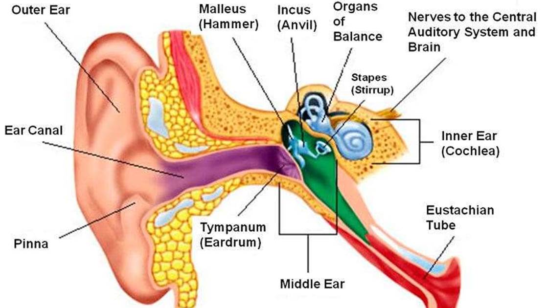
The following is a list of the steps that occur in the production of an auditory sensation. The ossicles auditory ossicles are the three smallest bones in the body the malleus the incus and the stapes. Week 3 Assignment 2 1.
Body short limb long limbprocess and lenticular process. Articular with one another via. The first is attached to the tympanic membrane the last to the circumference of the fenestra vestibuli the incus being placed between and connected to both by delicate articulations.
Sensory receptors activated impulse sent to CNS sensation perceptionFeedback. Of adult size at birth and subsequently alter little. Anatomy and Physiology questions and answers.
Ear space relative to that in the external auditory canal may occur for all of the following EXCEPT. The auditory nerve is also known as the _______ cranial nerve. 46 Identify the auditory ossicles.
All of the following are true about the middle ear EXCEPT. Movement of the tympanic membrane causes displacement of the malleus. Medium Study Objective 1.
Constriction of the pupil. It consists of the. The joints between ossicles are synovial.
Displacement of the stereocilia stimulates sensory neurons of the cochlear nerve. The sacrum and _____ comprise the sacral spine. Which of the following is a function of the axial skeleton.
Cochlea to the round window. Change in the curvature of the lens C. The incus anvil is the middle auditory ossicle.
First bone of the inner ear attaches to the tympanic membrane and the incus. The auditory tube connects the nasopharynx with the anterior wall. Provides an attachment for muscles that move the head neck and trunk c.
The pressure wave distorts the basilar membrane on its way to the round window. Mastoid antrum is an air sinus in the petrous part of temporal bone. Stapes The three ossicles in the middle ear are the malleus incus and stapes.
The auditory ossicles connect the. The middle-ear ossicle that rocks in the oval window to transmit the mechanical vibrations of the ossicles to the. The head lies in the epitympanic recess.
Adjustment to close-range vision involves all of the following except _____. Like the head of the malleus it sits in. All of the following are extraocular muscles that are responsible for the motion of the eyes except.
Its adult capacity is variable but on average is 1 mL with a. The ________ is a bony structure located between the cochlea and the three semicircular canals. All of the following are true about the middle ear EXCEPT.
Humans have _____ vision because of the overlap between the two visual fields. The malleus malleus or hammer consists of a head caput mallei neck collum mallei handle or manubrium manubrium mallei that ends as the spatulate process processus spatuliforms and two other processes. Malleus and incus are derived from.
SO 1741 Describe the anatomy of the structures in the three main. Cochlear duct spiral organ ossicles oval window auditory canal tympanic membrane fibers of cochlear nerve. Activity of the extrinsic eye muscles B.
All of the following statements accurately describe the auditory ossicles except They are located in the petrous part of the parietal bones. The facial nerve passes in a canal situated in the medial and anterior walls. The external ear includes all of the following except the.
Identify and apply the knowledge of all body systems their structure and functions and their common diseases symptoms and etiologies. The Auditory Ossicles - Human Anatomy. The chorda tympani nerve is related to the lateral wall.
Negative pressure in the middle-ear space relative to that in the external auditory canal may. The three auditory ossicles ossicula auditus or ossicula auditoria are the malleus the incus and the stapes see Fig. Aqueous humor Vitreous humor Perilymph O Endolymph.
See full answer below. The body of the incus articulates with the head of the malleus anterolaterally. Maxillary antrum orbit The tympanic cavity Tympanic or mastoid antrum auditory ear osicles and internal ear structures are all almost fully developed ie.
The correct answer is. Question 48 Correct Mark 100 out of 100 Flag question Question text Rods are more sensitive to light than cones and rods are important for color vision.

Auditory Ossicles Www Medicoapps Org
The Auditory Ossicles Smallest Bones In The Body Flashcards Quizlet

The Sequence Of Ear Ossicles From Outside Tympanum To Inside Is


0 Comments